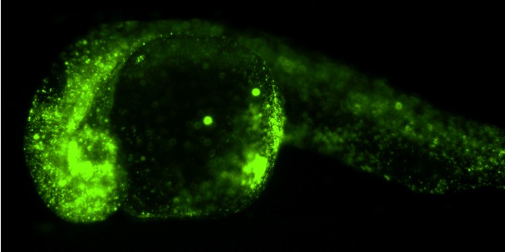
Researchers investigate mechanical features of cells
Cells form tissues or organs, migrate from place to place and in doing that their mechanical features and forces generated within them play a key role. Researchers at the Cells-in-Motion Cluster of Excellence at Münster University have now investigated the mechanical features of cells in living zebrafish embryos using the holographic optical tweezers-based method. In the process, cell biologists and physicists worked closely together. The light-based technology enabled them, for the first time, to manipulate several components in the cells simultaneously and carry out measurements. The study appears in the current issue of the Journal of Biophotonics (online).
In the Cells-in-Motion Cluster of Excellence, a team of cell biologists headed by Prof. Erez Raz observes primordial germ cells – i.e. the precursors of sperm and egg – as they travel around the developing zebrafish. Just like other cells, a germ cell moves forwards by deforming, with small blebs of the cell membrane forming in the direction it is moving in. Determining the mechanical properties of the cell and its surroundings is essential for understanding this process. These mechanical features are important not only during the development of organisms, but also in diseases – for example, when cancer cells travel and form metastases. “When a cell divides, or changes during an illness, its mechanical properties can change as well”, says Florian Hörner, a PhD student at the Institute of Cell Biology and the lead author of the study.
A group of researchers led by Prof. Cornelia Denz, a physicist, contributed a special method for investigating these physical properties – holographic optical tweezers. Optical tweezers are based on a focused laser beam, by means of which very small particles in a cell or tissue can be moved, deformed and held in place. The laser acts as "tweezers" made of light. In order to hold more than just one single particle in place, the researchers combined the optical tweezers principle with holographic methods. The hologram produced by the computer allows shaping the light to generate a large number of individual optical tweezers under the microscope.
To be able to examine cell properties during embryonic development, the researchers injected tiny plastic beads, one micrometre in size, into cells of zebrafish embryos at an early stage in their development. By means of the optical tweezers, the researchers pushed the beads and moved them within in the cell. Precise analysis of the movement of the beads in response to the push allowed the researchers to determine mechanical properties within the cells. In addition, employing the holographic tweezers it was possible to exert forces at different locations simultaneously – as if using several hands to manipulate the shape of structures within cells.
"We have found a good platform for looking into many different questions concerning the mechanical features of cells within a living embryo, without interfering with their development," says Florian Hörner. The researchers succeeded to establish a versatile method, which can be used in the future for similar studies involving other organisms. "Ultimately," says Prof. Erez Raz, "we want to be able to measure and manipulate biophysical cellular properties relevant for the migration of cells within live tissues".
The study originated in a research project funded by the Cells-in-Motion Cluster of Excellence. It also was supported by the Collaborative Research Centre TRR 61 ("Multilevel Molecular Assemblies – Structure, Dynamics and Function") at Münster University – funded by the German Research Foundation, as well as from the European Research Council and the German Academic Exchange Service.
Original publication:
Hörner F, Meissner R, Polali S, Pfeiffer J, Betz T, Denz C, Raz E. Holographic optical tweezers-based in vivo manipulations in zebrafish embryos. J Biophotonics 2017, DOI: 10.1002/jbio.201600226
