Introduction
The general problem of many types of analysis is the plenty of data in the opposite to the available time. To reduce analysis time and manpower, computers are needed. Visual information is a special type of data, for humans easy to recognise, for maschines only a 2-dimensional array containing grey levels. To find the desired information automatically, the images are analysed by Digital Image Processing.DNA-Analysis
One application is the analysis of chromosoms. Blood cells are irradiated by 60Co and the relation between dose and damage is researched. A probe of irradiated blood on an object slide is typically in the dimension 2cm x 3cm. The field of view during scanning this probe with a microscope with 100x objective is nearly 100mum x 100mum.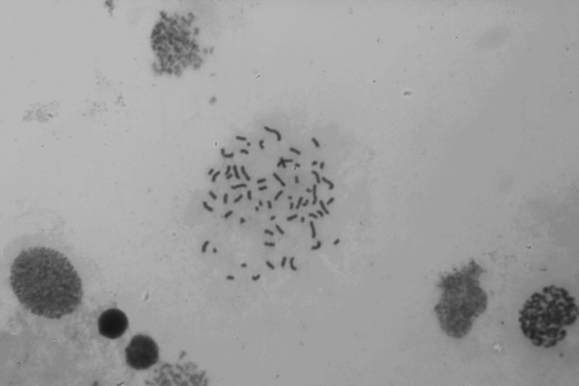
Chromosoms of a splashed blood cell. The big bubbles around are not-splashed cells.
The blood probe has to scanned completly, about nothing to overlook. The chromosomes have to be count; the normal, healthy cell has 46 chromsoms. This type of pattern recognicion and counting is a typical application for Image Processing. Couppled with motorised high-precition movement of the optical stage, the probe can be scanned systematically.

 is under construction.
Back to project Last modified Apr 1999
Nicola Bruski / bruski@uni-muenster.de